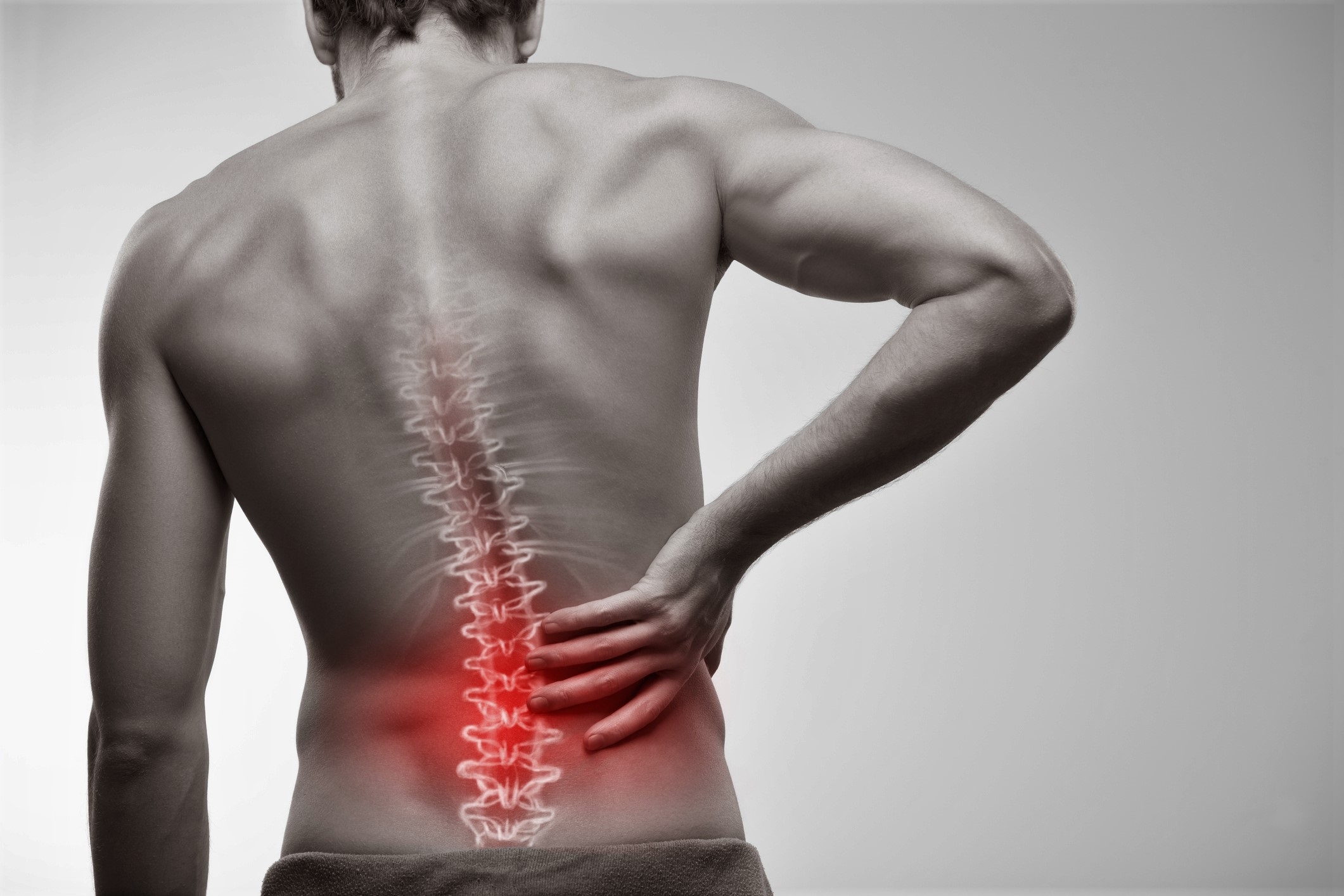
Understanding Sciatica and Radiculopathy: Causes, Symptoms, and Management
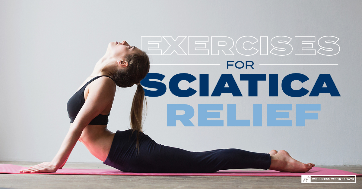
What Are the Top 10 Exercises for Sciatica Pain Relief?
How Does Proper Hydration Reduce Muscle Pain Naturally?
Are Mesh Back Chairs Better for Sciatica Pain Relief?
How Does Stretching Help in Easing Sciatica Discomfort?
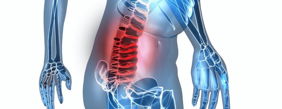
Nerve Root Compression: Understanding, Symptoms, and Effective Treatments
Understanding and Treating a Herniated Disc: Your Guide to Relief and Recovery
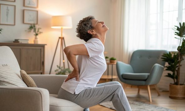
Lower Back Pain: What’s Really Going On and How You Can Feel Better
How Does Regular Exercise Help Alleviate Lower Back Pain?
How Does Aging Contribute to Chronic Lower Back Pain?
Are Core Strengthening Exercises Effective for Lower Back Pain Relief?
How Often Should You Exercise to Maintain Lower Back Health?
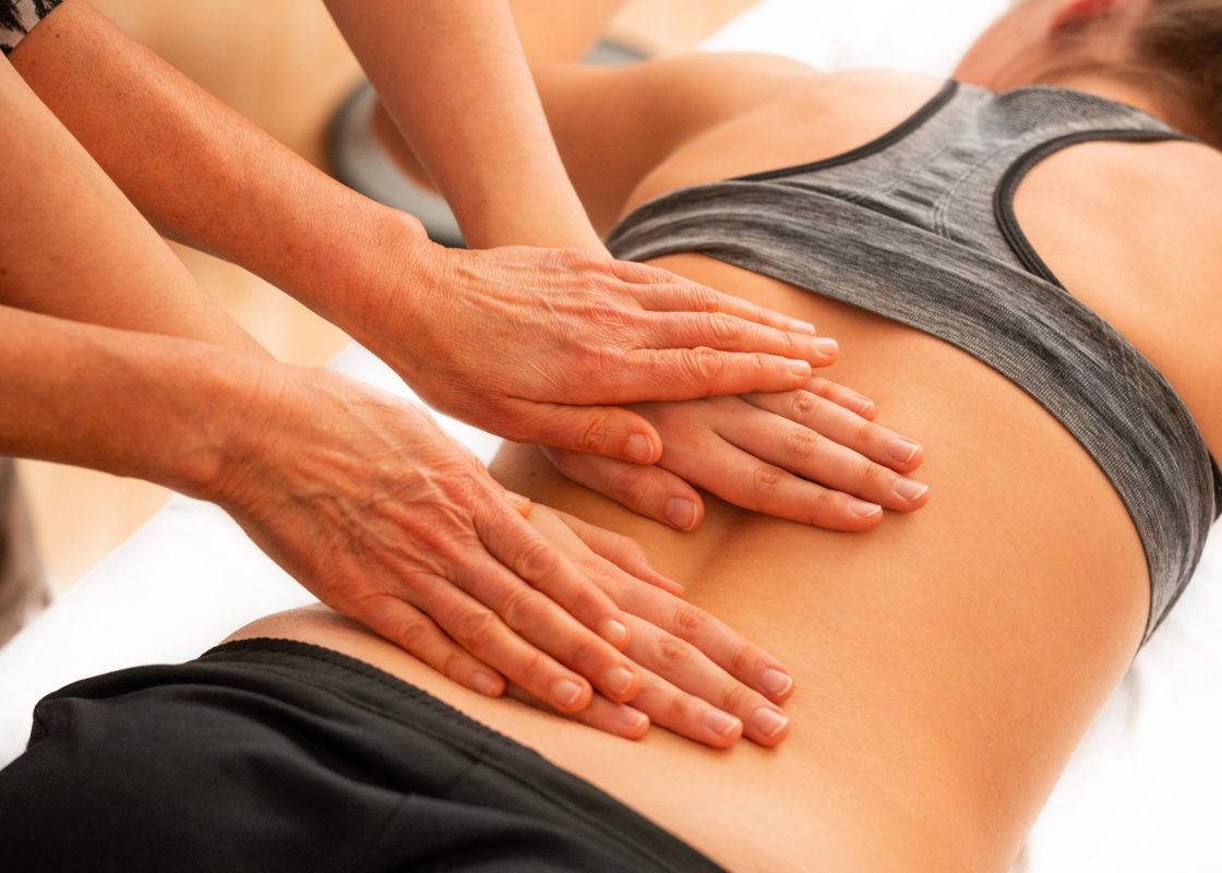
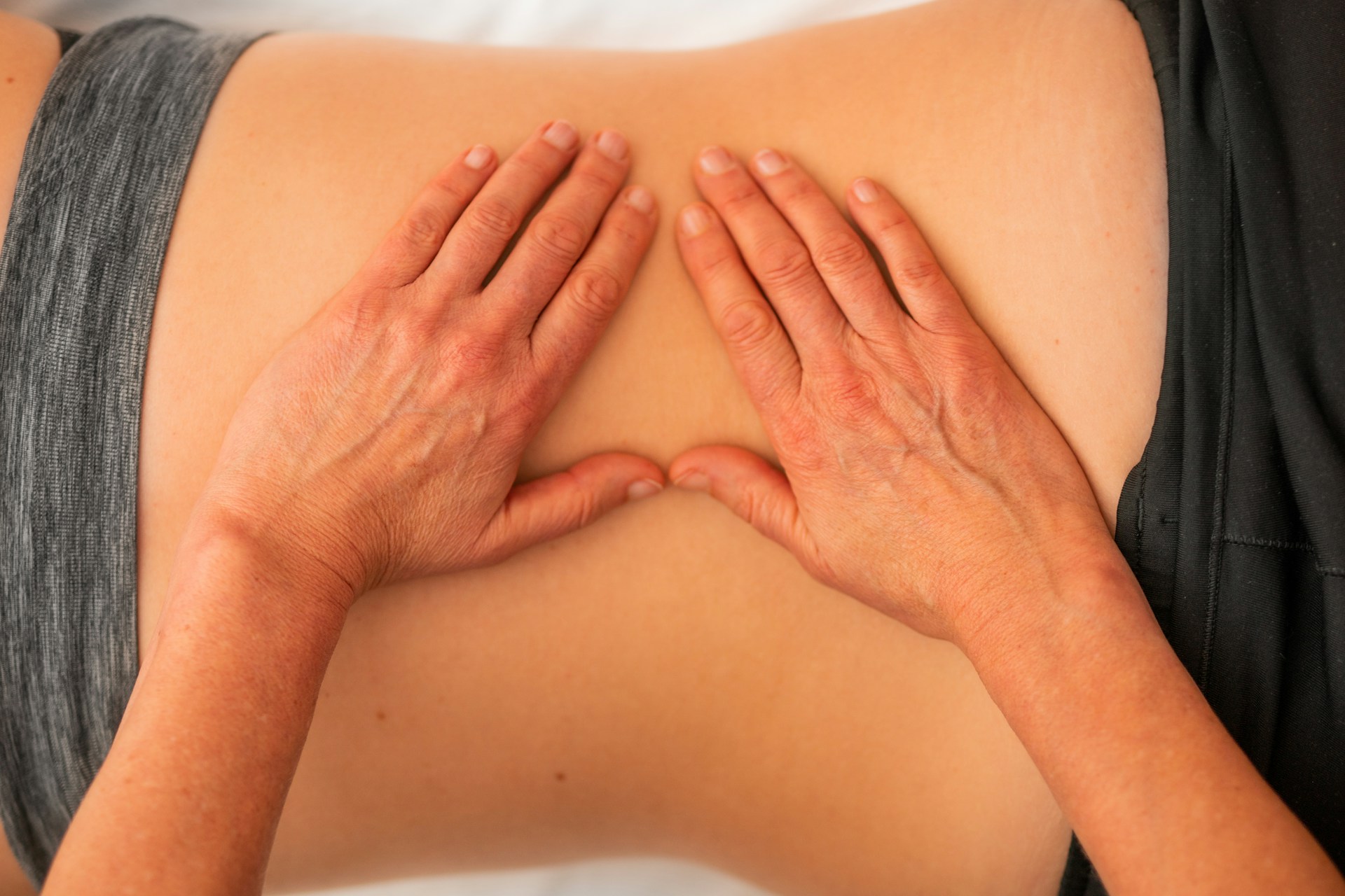
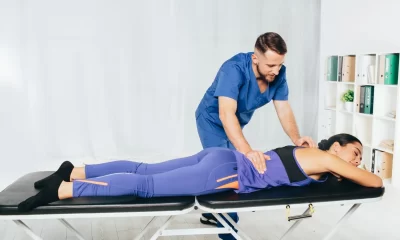
What Are the Phases of Chiropractic Care?
Why Do Chiropractors Require So Many Visits?
How Effective is Chiropractic Treatment?
What Does Chiropractic Care Do?
How Do Chiropractors Know Where to Crack?
How Does CBD Oil Make You Feel?
What Is CBD Oil Good For?
5 of the best CBD oil for anxiety
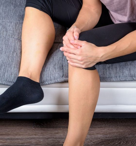
Leg pain affects millions of people worldwide and can significantly impact daily activities, mobility, and quality of life. While some...
Nerve pain can be debilitating and confusing, often manifesting in various forms. Among the most commonly discussed conditions are sciatica...